In February 2007, I participated in a voluntary study at the Institute of Cognitive Neuroscience in London. The study involved three 2-hour sessions consisting of different 10-minute tasks which were greatly varied (presumably to exercise different areas of your brain and to score you against several metrics).
The last session involved several MRI and fMRI scans while completing tasks in the noisy environment of the scanner. Thanks to a friend who worked at the Institute, I was able to secure a copy of the raw data and an application to turn it into images, some of which are displayed here.
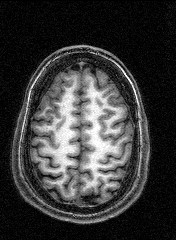
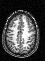
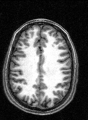
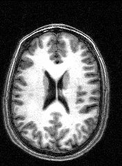
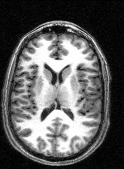
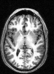
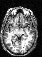
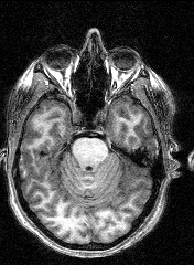
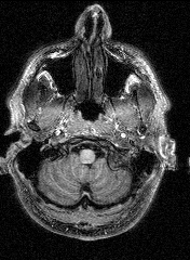
The images to the right are virtual 'slices', taken horizontally, starting from the top and working down. Cerebrospinal fluid is dark, the white matter appears light.
Medical images such as these are easy to come by on the internet, but having images of your own brain brings a humbling perspective.